CARDIOLOGY - ECG LIBRARY
Case 64
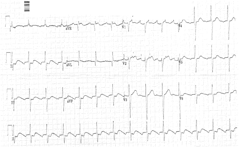
36F, Syncope. i) State rhythm and differential diagnosis. ii) Describe 3 abnormalities.
Expected Response
Narrow complex regular rhythm at a rate of 85
Flutter waves seen in V1 and V2
Likely atrial flutter with 2:1 or 3:1 block
Ddx Sinus rhythm although can’t see p waves
3 abnormalities on the ECG and explained how each may lead to syncope.
Long QT - >1/2 R-R interval – could have caused Torsades
Evidence of HOCM – ‘dagger-like’ Q waves in aVL and Leftward axis
Outflow tract obstruction or arrhythmia related to HOCM (this patient actually has HOCM)
Atrial flutter Could have caused syncope with 1:1 conduction and rapid ventricular response causing syncope
Flutter waves seen in V1 and V2
Likely atrial flutter with 2:1 or 3:1 block
Ddx Sinus rhythm although can’t see p waves
3 abnormalities on the ECG and explained how each may lead to syncope.
Long QT - >1/2 R-R interval – could have caused Torsades
Evidence of HOCM – ‘dagger-like’ Q waves in aVL and Leftward axis
Outflow tract obstruction or arrhythmia related to HOCM (this patient actually has HOCM)
Atrial flutter Could have caused syncope with 1:1 conduction and rapid ventricular response causing syncope