VASCULAR
AORTIC DISSECTION
Elderly hypertensive male
PromptLife threatening chest pain DDx
ResponseSTEMI (or NSTEMI)
Aortic dissection
PE - Massive PE
AAA rupture
Cardiac arrhythmia
Boerhaave syndrome/esophageal rupture
Aortic dissection
PE - Massive PE
AAA rupture
Cardiac arrhythmia
Boerhaave syndrome/esophageal rupture
PromptRisk factors for aortic dissection
ResponseConnective tissue disease (Ehlers Danlos, Marfan’s)
Family history of aortic disease
Htn
Bicuspid aortic valves
Coarctation of the aorta, Aortic stensosis
Pre-existing aneurysmal dilation
Old + male
Iatrogenic - recent angioplasty/bypass/valve surgery
Family history of aortic disease
Htn
Bicuspid aortic valves
Coarctation of the aorta, Aortic stensosis
Pre-existing aneurysmal dilation
Old + male
Iatrogenic - recent angioplasty/bypass/valve surgery
PromptExamination findings in aortic dissection
ResponseMurmur of AR
Signs of pericardial effusion
Evidence of branch dissection/perfusion deficit
Patient distress - diaphoresis, pallor, tachycardia
Other - severe hypertension, risk factors (eg: marfan’s syndrome)
Signs of pericardial effusion
Evidence of branch dissection/perfusion deficit
- Carotid/vertebral = focal neurology
- BP difference between arms (Brachiocephalic or Subclavian involvement) but not specific
- Spinal level from spinal infarct
Patient distress - diaphoresis, pallor, tachycardia
Other - severe hypertension, risk factors (eg: marfan’s syndrome)
PromptMost useful initial investigations for chest pain and collapse with suspected dissection
ResponseECG
- ? Coronary artery involvement/secondary myocardial ischaemia
- Only use if unable to perform CT.
- TTE not very sensitive - unable to visualise much of aorta (OK for ascending)
- TOE better but less available/tolerated.
- Best for tamponade and valve involvement
- Preferable study - definitive diagnosis, aids in repair planning. Contraindicated with allergy to contrast, inability to tolerate CT (Haemodynamics, laying flat). Relative contraindication in renal failure. If CTPA in stem ask for a CT aorta
- Can’t assess aortic incompetence
PromptECG findings
ResponseSTEMI mimic - inferior MI
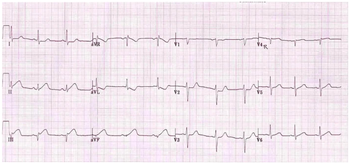
PromptDDx of wide mediastinum on cxr
ResponseAP film + bad magnification effect
Unfolded aortic arch
Thoracic aortic aneurysm
Paravertebral Mass
Oesophageal dilation
Mediastinal lymphadenopathy
Unfolded aortic arch
Thoracic aortic aneurysm
Paravertebral Mass
Oesophageal dilation
Mediastinal lymphadenopathy
Prompt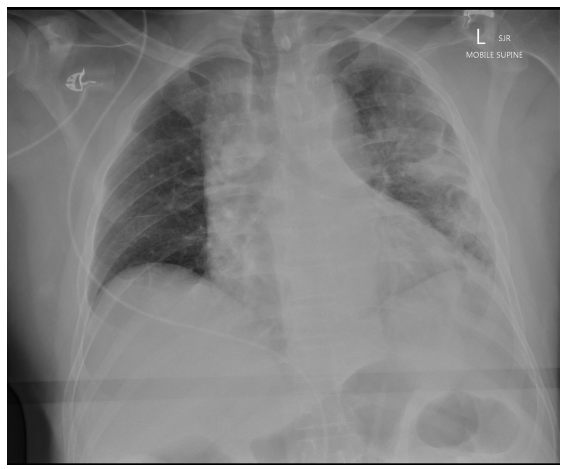
ResponseWidened mediastinum despite AP supine view (normal <8cm on PA film)
Loss of aortic knob contour
Patchy opacities L lung field ?Infective/aspiration
Loss of L costophrenic angle - possible small effusion
Likely diagnosis - aortic dissection
Loss of aortic knob contour
Patchy opacities L lung field ?Infective/aspiration
Loss of L costophrenic angle - possible small effusion
Likely diagnosis - aortic dissection
PromptCXR findings concerning for dissection
ResponseWidening of the superior mediastinum
Dilation of the aortic arch
Chang in the configuration of the aorta on successive CXR
Obliteration of the aortic knob (Notch)
Double density of aorta (Suggests true and false lumen)
Localised prominence along aortic contour
Disparity of caliber between descending and ascending aorta
Calcium sign - >6mm between intimal calcium and shadow of outer aortic wall
Displacement of trachea to right (Or NG tube)
Distortion of left main stem bronchus
Pleural effusion
Cardiomegaly
Dilation of the aortic arch
Chang in the configuration of the aorta on successive CXR
Obliteration of the aortic knob (Notch)
Double density of aorta (Suggests true and false lumen)
Localised prominence along aortic contour
Disparity of caliber between descending and ascending aorta
Calcium sign - >6mm between intimal calcium and shadow of outer aortic wall
Displacement of trachea to right (Or NG tube)
Distortion of left main stem bronchus
Pleural effusion
Cardiomegaly
PromptBedside US (MI vs Dissection)
ResponseMI:
- Regional wall motion abnormality/hypokinesis
- Pericardial effusion +RV collapse + dilated IVC (pericardial tamponade)
- AV incompetence/regurgitation
- Intimal flap (Sternal notch view)
Prompt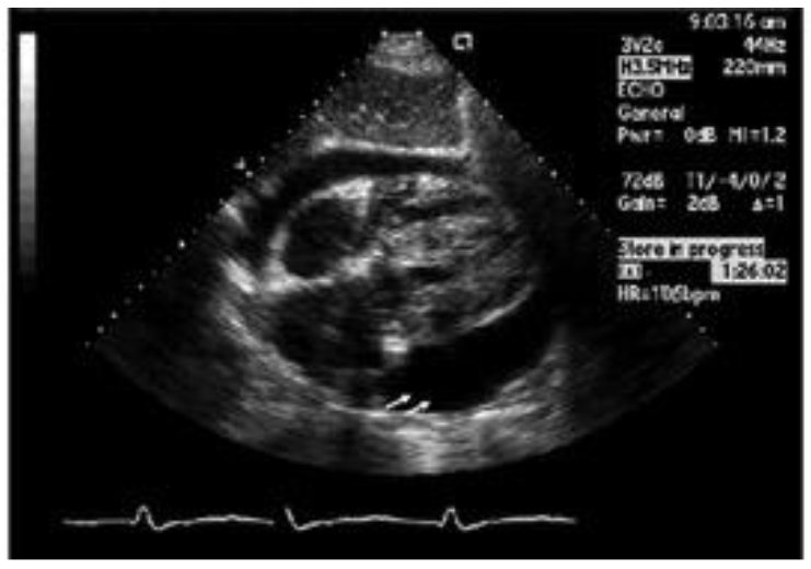
ResponseAbnormality: Large pericardial effusion
Diagnosis: Thoracic aortic dissection with pericardial effusion, impending tamponade
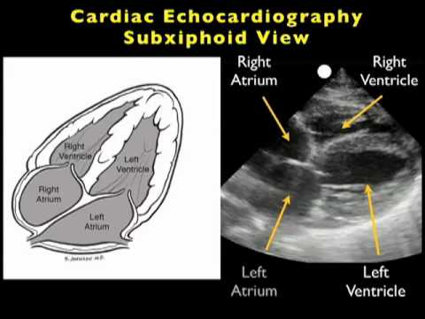
Diagnosis: Thoracic aortic dissection with pericardial effusion, impending tamponade
PromptThis bedside ultrasound taken as pt arrests
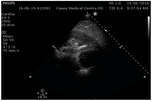
ResponseLarge pericardial effusion
Collapsed RA and RV
Non collapsed tricuspid annulus
Diagnosis = Type A thoracic aortic disection
Collapsed RA and RV
Non collapsed tricuspid annulus
Diagnosis = Type A thoracic aortic disection
Prompt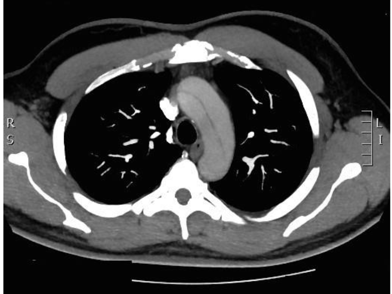
Response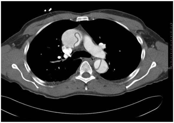
Type A aortic dissection
Note no points if don’t say Type A
Type A aortic dissection
Note no points if don’t say Type A
PromptD dimer use in evaluation of aortic dissection
ResponseADviSED Trial 2018 - only prospective evaluation of comibingin clinical risk stratification with D dimer. Multi centre 1850 patients. 98.8% sensitive to exclude dissection (8 patients with negative dimer had AAS)
Most authors suggest this holds promise but needs external validation for reproducibility
Most authors suggest this holds promise but needs external validation for reproducibility
PromptManagement options for dissection
ResponseOperative - urgent cardiothroacic surgery to replace ascending aorta - may need urgent pericardiocentesis if becomes haemodynamically unstable
Non operative - Palliative approach (mortality 1% per hour, >90% untreated)
Non operative - Palliative approach (mortality 1% per hour, >90% untreated)
PromptManagement of dissection
ResponseManage in resuscitation bay. Management is time critical
2 large bore IVC’s, bloods including G+H
IV analgesia (Fentany 25 mics titrated to comfortl/morphine)
Chronotherapy HR ~60 (Rate control first) - Reduces shear forces
Urgent transfer to cardiothoracic centre for CT +/- OT
Reversal of anticoagulation
Endpoints: SBP <120, HR<60, Pain well controlled
2 large bore IVC’s, bloods including G+H
IV analgesia (Fentany 25 mics titrated to comfortl/morphine)
Chronotherapy HR ~60 (Rate control first) - Reduces shear forces
- IV rate control (Beta blocker, metoprolol, esmolol) aim HR ~60
- Esmolol seems best but Metoprolol 2.5mg IV + repeat = familiar
- IV BP control (GTN, SNP, Hydralazine) - aim SBP <140
- GTN 5-50 mics/min (50mg in 500 mL D5W, start 3 mL/h)
Urgent transfer to cardiothoracic centre for CT +/- OT
Reversal of anticoagulation
Endpoints: SBP <120, HR<60, Pain well controlled
PromptDrugs to reduce shear forces - pros and cons
ResponseEsmolol (2.5g in 250 mL, start at 20 mL/h - 50-200 mics/kg/min)
- Rapid onset, titratable, short action of duration - Can be switched off if hypotension/bronchospasm. Clearance not affected by hepatic/renal function
- Unfamilliary to staff, need continuous infusion, continuous monitoring
- IV available, familiar to staff
- Not so titratable, long duration of action if things go bad
- Available IV, use if beta blockers contraindicated (Asthma etc.). Intermediate duration of action
- Reduce adrenergic outflow and therefore contractility and HR
PromptComplications of Dissection (and causes of hypotension)
ResponsePericardial tamponade from pericardial extension - on bedside echo
Aortic rupture - haemothorax on CXR/eFAST
Stroke
Severe AR - Aortic valve incompetence from aortic root dissection and pump failure - murmur with pulmonary congestion and empty pericardium.
MI from aortic root involvement - STEMI on ECG
(Other organ ischaemia due to dissected branch)
Iatrogenic hypotension from excess BP/HR management
Branch vessel dissection - pseudohypotension (ie: dissected off circulation to the arm the bp cuff is on) - check other limbs
Aortic rupture - haemothorax on CXR/eFAST
Stroke
Severe AR - Aortic valve incompetence from aortic root dissection and pump failure - murmur with pulmonary congestion and empty pericardium.
MI from aortic root involvement - STEMI on ECG
(Other organ ischaemia due to dissected branch)
Iatrogenic hypotension from excess BP/HR management
Branch vessel dissection - pseudohypotension (ie: dissected off circulation to the arm the bp cuff is on) - check other limbs
PromptClassification of Dissection
ResponseStanford
A = Ascending +/- descending
B = Descending aorta only (distal to origin of left subclavian)
De Bakey
I = Ascending + Descending
II = Ascending only
III = Descending only. A = thoracic, B - extends to abdominal aorta
A = Ascending +/- descending
B = Descending aorta only (distal to origin of left subclavian)
De Bakey
I = Ascending + Descending
II = Ascending only
III = Descending only. A = thoracic, B - extends to abdominal aorta
PromptUS use in management of tamponade
ResponseUS guided vs landmarks
Identify biggest pocket of fluid
Use of contrast (adgitated saline)/location of needle
Identify the cause of tamponade - ie Ventricular rupture/dissection to direct specific treatment.
Identify biggest pocket of fluid
Use of contrast (adgitated saline)/location of needle
Identify the cause of tamponade - ie Ventricular rupture/dissection to direct specific treatment.