ULTRASOUND, ECHO AND FAST
eFAST
PromptRole of eFAST in haemodynamically stable blunt trauma patients
ResponseClear role for FAST in unstable blunt trauma (guides decisions about going to OT…)
In stable patients, sensitivity of FAST in detecting significant injury is poor (< 50% - i.e. lots of false negatives – doesn’t “rule out” the disease)
In particular misses hollow viscus & retroperitoneal injury and doesn’t assess solid organ injury (with view to non-operative Mx)
There is no evidence base to recommend FAST in this context. Decisions about CT scan/laparotomy/observation need to be based on other patient findings
May be useful for clinician practice and possibly a role for serial FAST in a stable patient admitted for observation
In stable patients, sensitivity of FAST in detecting significant injury is poor (< 50% - i.e. lots of false negatives – doesn’t “rule out” the disease)
In particular misses hollow viscus & retroperitoneal injury and doesn’t assess solid organ injury (with view to non-operative Mx)
There is no evidence base to recommend FAST in this context. Decisions about CT scan/laparotomy/observation need to be based on other patient findings
May be useful for clinician practice and possibly a role for serial FAST in a stable patient admitted for observation
PromptPro’s of eFAST
ResponseSensitive + specific for free fluid
Rapid at bedside
Expedites transfer to theatre
No radiation exposure (marks probably if 5 yo, not if 80yo)
Non invasive
Repeatable
Rapid at bedside
Expedites transfer to theatre
No radiation exposure (marks probably if 5 yo, not if 80yo)
Non invasive
Repeatable
PromptPitfalls/limitations of FAST Ultrasound in Trauma
ResponseOperator dependent
Technical difficulties – obesity, subcutaneous air
Requires > 250 mL blood to collect to become positive
Doesn’t look at retroperitoneal space, hollow viscera or solid organs
Role in pelvic trauma unclear
Doesn’t identify source of bleeding
Doesn’t distinguish other sources of IP fluid (peritoneal dialysis, ascites, ruptured ovarian cyst etc
False positive unnecessary further imaging/operation
Opportunity cost, in terms of staffing resources and time for other procedures ie. People dicking around with the probe instead of getting on with definitive care
Trauma patients non-fasted and limitations with bowel gas
Evidence for use in Paediatric population limited and due to primarily conservative approach in kids for blunt trauma - limited utility in decision
making.
Technical difficulties – obesity, subcutaneous air
Requires > 250 mL blood to collect to become positive
Doesn’t look at retroperitoneal space, hollow viscera or solid organs
Role in pelvic trauma unclear
Doesn’t identify source of bleeding
Doesn’t distinguish other sources of IP fluid (peritoneal dialysis, ascites, ruptured ovarian cyst etc
False positive unnecessary further imaging/operation
Opportunity cost, in terms of staffing resources and time for other procedures ie. People dicking around with the probe instead of getting on with definitive care
Trauma patients non-fasted and limitations with bowel gas
Evidence for use in Paediatric population limited and due to primarily conservative approach in kids for blunt trauma - limited utility in decision
making.
PromptList reasons for a false eFAST scan in abdominal trauma
Responsepelvic / encapsulated /retroperitoneal
small volume ie < 100 ml
operator ability
patient bowel gas
patient obesity
small volume ie < 100 ml
operator ability
patient bowel gas
patient obesity
Prompt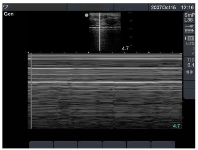
ResponseStratosphere or Barcode sign (Evidence of lack of pleural sliding)
R sided Tension pneumothorax leading to obstructive shock
R sided Tension pneumothorax leading to obstructive shock
PromptRUQ image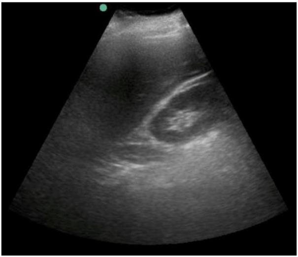
ResponsePossible hypoechoic stripe adjacent to kidney – wedge-shaped and bounded by echoic lines
likely represents perinephric fat rather than free fluid. (1 mark for adequate description, 1 mark for correct interpretation)
likely represents perinephric fat rather than free fluid. (1 mark for adequate description, 1 mark for correct interpretation)
Prompt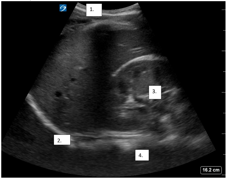
Response1. Skin/subcutaneous tissues
2. Diaphragm
3. R kidney
4. Spine/Vertebrae
2. Diaphragm
3. R kidney
4. Spine/Vertebrae
Prompt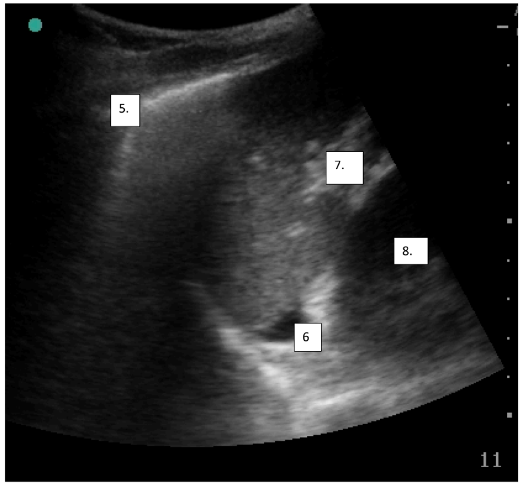
Response5. Lung point/edge
6. Free fluid
7. Splenic hilum
8. L kidney
Positive FAST - free fluid in pelvic and splenorenal view
6. Free fluid
7. Splenic hilum
8. L kidney
Positive FAST - free fluid in pelvic and splenorenal view
Prompt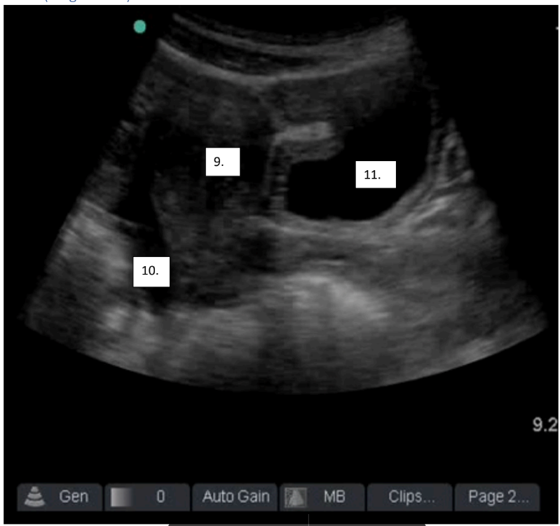
Response9. Uterus
10. Free fluid
11. Bladder
Positive FAST - free fluid in pelvic and splenorenal view
10. Free fluid
11. Bladder
Positive FAST - free fluid in pelvic and splenorenal view
Prompt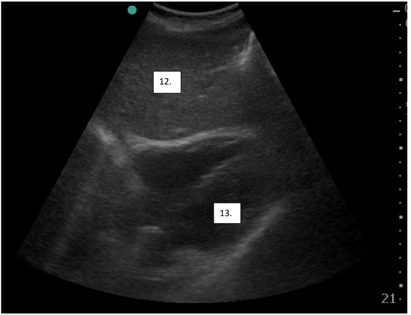
Response12. Liver
13. L ventricle
13. L ventricle
PromptHow would you make this image better?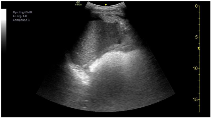
Responsedecrease gain
decrease depth
decrease depth
PromptShocked trauma patient RUQ
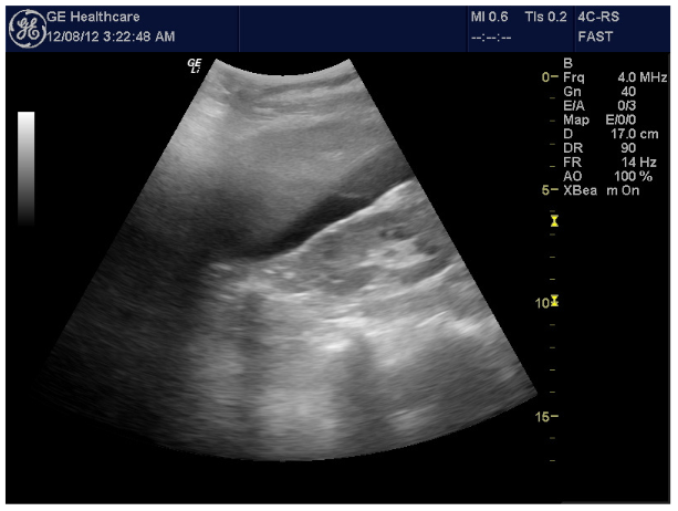
ResponseModerate/large amount of fluid in Morrison’s pouch
Interpretation = intraperitoneal blood
Interpretation = intraperitoneal blood
PromptShocked trauma patient, RUQ
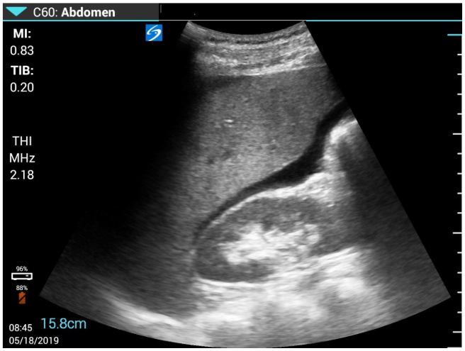
ResponseIntra-abdominal injury with free intraperitoneal fluid
= Strongly positive FAST scan demonstrating free fluid in Morrisons pouch
= Strongly positive FAST scan demonstrating free fluid in Morrisons pouch