TRAUMA - ADULT
CHEST - BLUNT
Much of this overlaps from penetrating.
Prompt27M, high speed MVA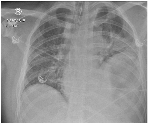
ResponseCXR Findings:
- ETT just above the level of his clavicles
- NG tube in situ that migrates below the level of the right hemidiaphragm and curls around to the left above the level of the right hemidiaphragm
- Deviation of the NG tube to the right
- Elevation of the left hemidiaphragm
- Fracture left scapula(subtle)
- Left lung basal collapse/consolidation
- Left apical capping
- Widened mediastinum
- Aortic dissection/transection
- Diaphragm rupture
- Cardiac injury(blunt)
- Lung injury-contusion/laceration
- Spleen injury
- Fractures- scapula, ribs
PromptList investigations in above patient in order of priority
ResponseEFAST
- assess for pericardial tamponade, assess for Type A dissection and assess for abdominal FF/PTx/HTx
- FBC, G+H, coag - suspected haemorrhage and coagulopathy of trauma
- given mechanism to assess for aortic injury, significant solid organ injury/abdominopelvic bleeding
- assess for shock - BE, lactate and response to Rx
Prompt40 M, High speed MVA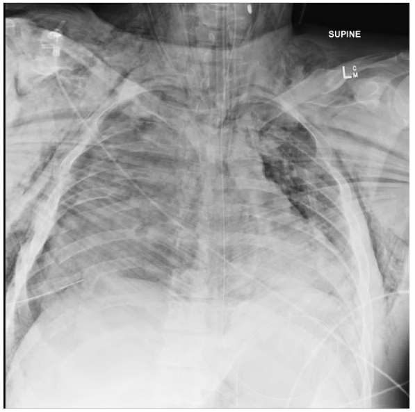
ResponseMultiple displaced Rib Fractures – displaced rib fractures seen posteriorly
Rt TENSION Pneumothorax – expansion Rt hemithorax, deviation NGT to left, visible lung edge, subcut emphysema
Rt Pulmonary Contusion – Increased opacification throughout Rt lung field
Lt Pneumothorax – subcutaneous emphysema
Left Clavicle fracture – comminution of mid-clavicle
Measures to improve oxygenation
Rt TENSION Pneumothorax – expansion Rt hemithorax, deviation NGT to left, visible lung edge, subcut emphysema
Rt Pulmonary Contusion – Increased opacification throughout Rt lung field
Lt Pneumothorax – subcutaneous emphysema
Left Clavicle fracture – comminution of mid-clavicle
Measures to improve oxygenation
- Rt thoracostomy tube
- Lt Thoracostomy tube
- Increase FiO2
- Increase PEEP
Prompt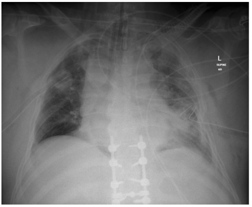
ResponseWidened mediastinum
Opacification both lung fields L > R
L rib #s (2-8)
L subcutaneous emphysema
N.B Intubated is not an acceptable answer as the ETT is adequately positioned. Consider accepting L ICC malpositioned or kinked as a possible abnormality.
Opacification both lung fields L > R
L rib #s (2-8)
L subcutaneous emphysema
N.B Intubated is not an acceptable answer as the ETT is adequately positioned. Consider accepting L ICC malpositioned or kinked as a possible abnormality.
PromptCauses for difficult ventilation in blunt chest trauma
Response
- ETT blocked = Hand ventilate with BVM, suction ETT, check position of ETT, replace ETT if necessary
- Tension Pneumothorax/Haemothorax, R ICC- drainage port borderline outside, the chest wall (progression of PTx) L ICC- kinked inside pleural cavity (progression of PTx) = Replace R side ICC if no swing noted in canister. Remove and replace ICC on L side.
- Pulmonary contusion = Increase PEEP and trial low volume/high rate ventilation
- Incorrect position of ETT eg migration into R main bronchus = Check ETT position & pull back to ensure ventilating both lungs
- Bronchospasm = Nebulised bronchodilators down ETT
PromptMVA - List 3 injuries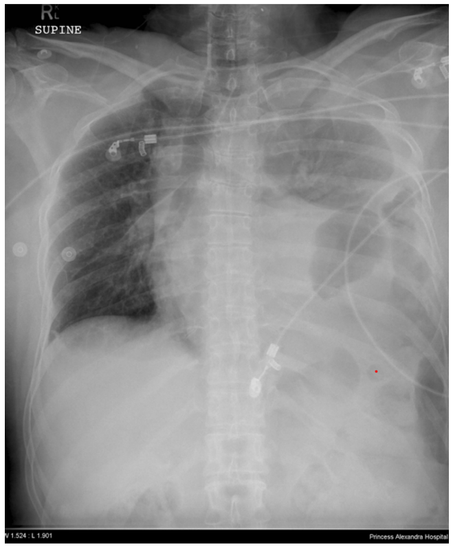
ResponseDiaphragmatic Rupture: NGT insertion, referral for operative repair
Left Haemothorax; ICC (careful insertion given diaphragmatic injury)
Multiple Lt rib fractures: analgesia, PCA, ?intercostal blocks
Left Haemothorax; ICC (careful insertion given diaphragmatic injury)
Multiple Lt rib fractures: analgesia, PCA, ?intercostal blocks
Prompt10m Fall, Main pathology + reasoning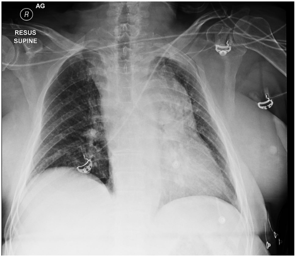
ResponseMain pathology: Mediastinal haematoma – likely great vessel injury
Reasoning
Wide mediastinum
Loss of aortic knob / Loss aorto-pulmonary window
Depressed left main bronchus
Widened right paratracheal stripe
Left apical pleural cap
Reasoning
Wide mediastinum
Loss of aortic knob / Loss aorto-pulmonary window
Depressed left main bronchus
Widened right paratracheal stripe
Left apical pleural cap
Prompt62M low speed MBA
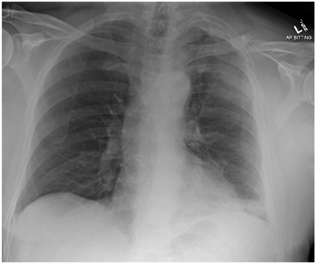
ResponseMultiple left-sided rib fractures including displaced fractures of 5th and 6th Ribs posterolaterally.
Comminuted # of the mid-third of the left clavicle with 13mm craniocaudal offset
Patchy airspace opacification at the left lower lobe – likely contusion.
No significant haemo- or pneumothorax
No subcutaneous emphysema
Cardiac contour appears normal
Further investigations (List + justify)
CT chest - Further define injuries seen and evaluate for subtle PTx, HTx, mediastinal haematoma, radiological flail.
ECG - Cardiac contusion (dysrhythmia or ST segment changes)
FBE, UEG, LFT, lipase, G&H, coags - Evaluate Hb (and baseline), G&H in case of transfusion
Blood gas - Early signs of hypoventilation secondary to rib # pain (raised pCO2).
Comminuted # of the mid-third of the left clavicle with 13mm craniocaudal offset
Patchy airspace opacification at the left lower lobe – likely contusion.
No significant haemo- or pneumothorax
No subcutaneous emphysema
Cardiac contour appears normal
Further investigations (List + justify)
CT chest - Further define injuries seen and evaluate for subtle PTx, HTx, mediastinal haematoma, radiological flail.
ECG - Cardiac contusion (dysrhythmia or ST segment changes)
FBE, UEG, LFT, lipase, G&H, coags - Evaluate Hb (and baseline), G&H in case of transfusion
Blood gas - Early signs of hypoventilation secondary to rib # pain (raised pCO2).
Prompt45F Fall 2m onto L chest
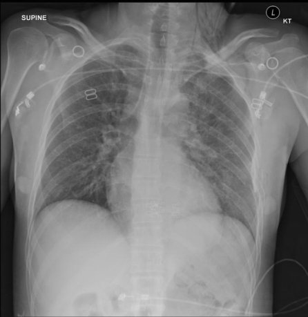
ResponseLeft pneumothorax – subcutaneous emphysema
Multiple rib fractures – left lateral displaced fractures visible
Left Haemothorax -veiling of left hemithorax (subtle)
Multiple rib fractures – left lateral displaced fractures visible
Left Haemothorax -veiling of left hemithorax (subtle)
PromptUnstable hypoxic MVA
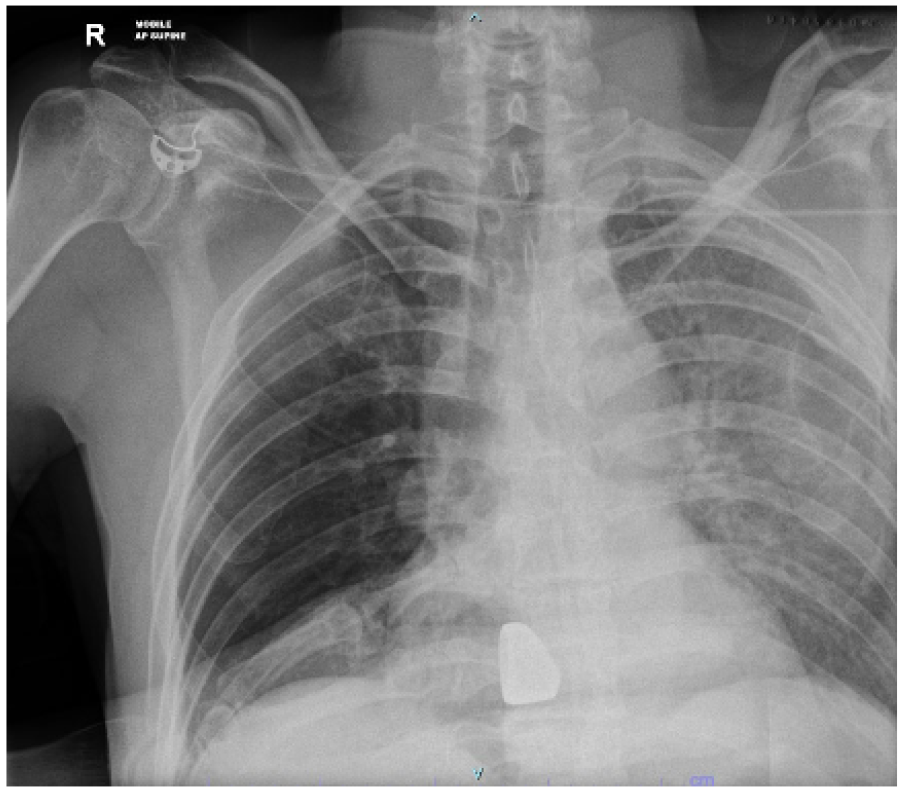
ResponseBlunt aortic injury - widened mediastinum/abnormal aortic contour, perhaps L apical capping
L sided haemothorax - diffuse L sided opacification. ? would also accept contusion, though heart border v. well defined going against this.
Ring on finger across chest
immediate emergency department management?
Finger thoracostomy +/- ICC
L sided haemothorax - diffuse L sided opacification. ? would also accept contusion, though heart border v. well defined going against this.
Ring on finger across chest
immediate emergency department management?
Finger thoracostomy +/- ICC
Prompt32M, MVA. tubed prehospital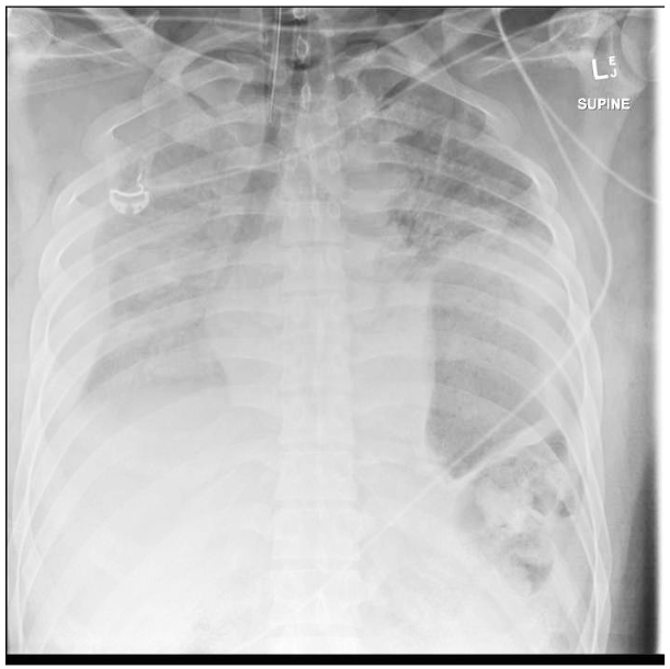
ResponseRight large haemothorax
- Veiled opacification of right hemithorax
- Substantial rim of blood around outside of lung edge
- Stomach clearly evident superiorly displaced in thorax
- Loss of diaphragmatic outline
- Opacification of visible left lung in upper zone
Prompt25M, MVA, Tubed prehospital. Name pathology and radiographic feature
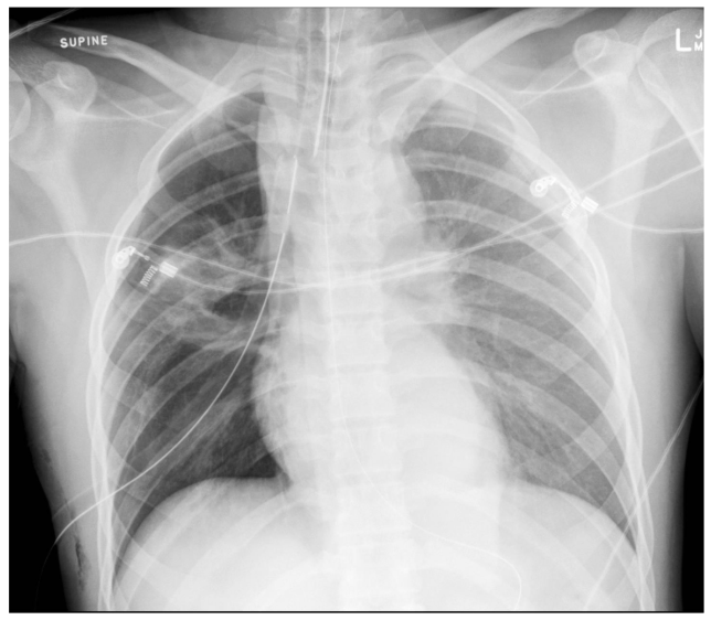
ResponseLeft haemothorax – veiled hemithorax, pleural cap
Mediastinal haematoma – wide mediastinum, rightward displacement NGT, paratracheal stripe, depressed left main bronchus
Right pulmonary contusion – right midzone opacities/consolidation
Right PTX – lucent right hemithorax, subcut emphysema
Mediastinal haematoma – wide mediastinum, rightward displacement NGT, paratracheal stripe, depressed left main bronchus
Right pulmonary contusion – right midzone opacities/consolidation
Right PTX – lucent right hemithorax, subcut emphysema
Prompt25F, MVA high speed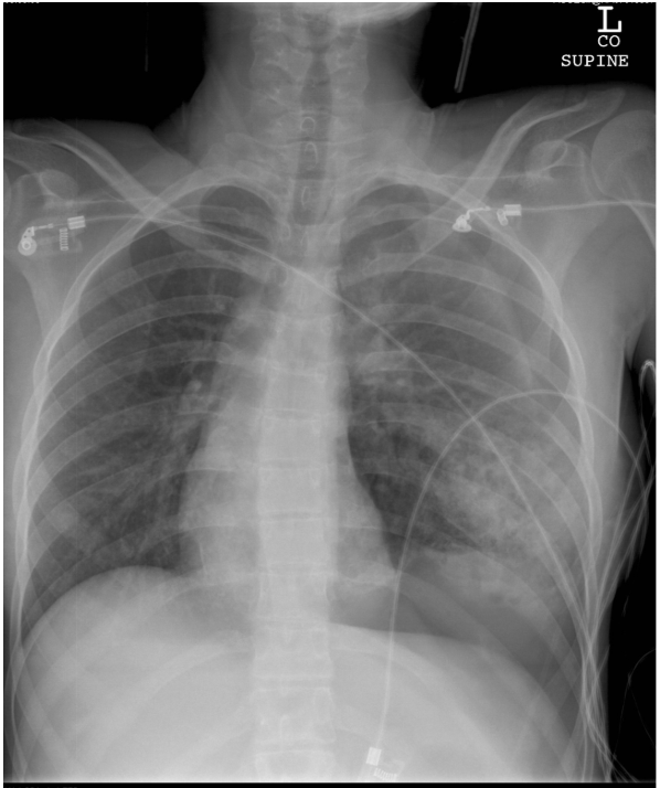
ResponseLeft tension pneumothorax
- Lung edge visible left upper lobe approx. 2cm in from chest wall
- Deep sulcus sign – basal air accumulation
- Mediastinal shift to right
- Definitive treatment = 32 Fr left ICC
Prompt38F, Fall 10m - Abnormal findings
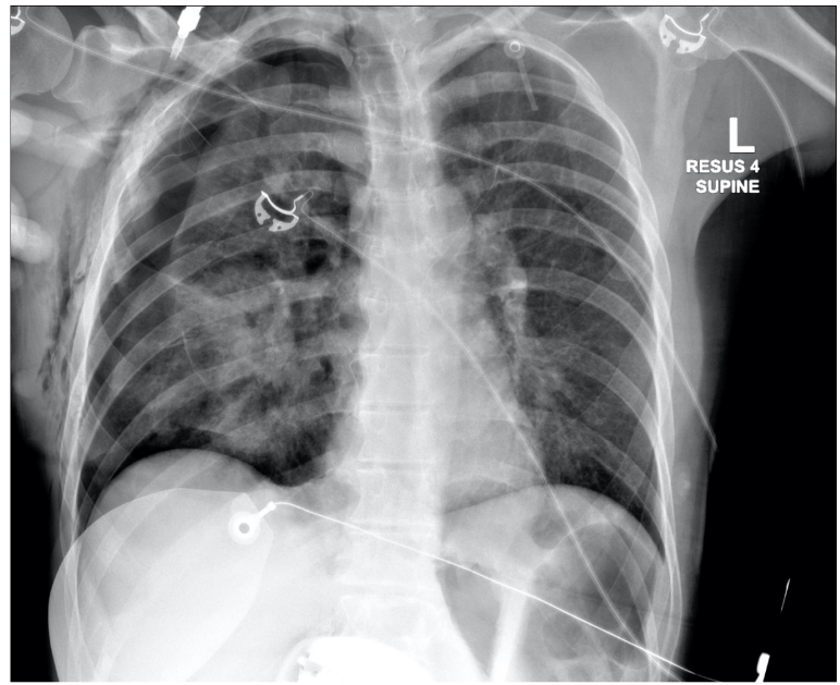
ResponseRight sided pneumothorax with mediastinal shift indicating signs of tension
Multiple right sided rib fractures
Needle from chest decompression attempt in situ right side of chest, possibly extra-thoracic
Extensive subcutaneous emphysema right chest wall
Small heart size – possibly from tension or massive blood loss
Increased opacity of collapse lung suggestion lung contusion
Multiple right sided rib fractures
Needle from chest decompression attempt in situ right side of chest, possibly extra-thoracic
Extensive subcutaneous emphysema right chest wall
Small heart size – possibly from tension or massive blood loss
Increased opacity of collapse lung suggestion lung contusion
PromptBenefits of performing a CT in chest trauma
ResponseIdentify ongoing source of bleeding – blush on CTA - eg intercostal vascular focus
Identify other associated intra-thoracic injuries ie contusions, pulmonary lacerations
Quantify PTX to aid decision making around need for/ type of ICC
Quantify size of haemothorax and need for intervention
Identify evidence of radiologic flail and inform possible need for rib fixation
Exclude thoracic spine fracture
Identify other associated intra-thoracic injuries ie contusions, pulmonary lacerations
Quantify PTX to aid decision making around need for/ type of ICC
Quantify size of haemothorax and need for intervention
Identify evidence of radiologic flail and inform possible need for rib fixation
Exclude thoracic spine fracture
PromptComplications of rib fractures
ResponseAtelectasis
Hypostatic pneumonia
Empyema
Progressive pneumothorax
Pleural fibrosis – restrictive lung disease
Hypostatic pneumonia
Empyema
Progressive pneumothorax
Pleural fibrosis – restrictive lung disease
PromptAnalgesia options for rib fractures
ResponseIV opiate ie fentanyl 50mcg repeated to comfort
PCA ie fentanyl or oxydodone ie oxycodone 1-2mg with 5 min lockout
Ketamine infusion
Intercostal nerve block under USS guidance
Serratus Anterior plane block under ultrasound
Oral drugs – paracetamol, ibuprofen, oxycodone doses
PCA ie fentanyl or oxydodone ie oxycodone 1-2mg with 5 min lockout
Ketamine infusion
Intercostal nerve block under USS guidance
Serratus Anterior plane block under ultrasound
Oral drugs – paracetamol, ibuprofen, oxycodone doses
PromptIndications for immediate operative management in isolated chest trauma
ResponseInitial dump >1500mL from ICC
Refractory shock
Tamponade
Tension pneumomediastinum
Refractory shock
Tamponade
Tension pneumomediastinum
PromptCauses of agitation in trauma patients
ResponseHypoxia
Hypovolaemia - ie shock. Only one point for blood loss.
Drugs - taken prior to arrival or during ICC insertion
Intracranial pathology
Hypercapnoea Hypoglycaemia
Hypovolaemia - ie shock. Only one point for blood loss.
Drugs - taken prior to arrival or during ICC insertion
Intracranial pathology
Hypercapnoea Hypoglycaemia
PromptModifications to RSI in unstable chest trauma patient
ResponseDecreased dose of induction agent – Ketamine 0.5mg/kg to mitigate haemodynamic depression
Hi-Flow Oxygen throughout induction – NP O2 @ 15lpm + BVM @ 15lpm through induction/apnoea
Avoid apnoea – bag through induction to avoid acidosis and hypoxia
Fluid load to counter vasodilation – 1u PRBCs during induction
Hi-Flow Oxygen throughout induction – NP O2 @ 15lpm + BVM @ 15lpm through induction/apnoea
Avoid apnoea – bag through induction to avoid acidosis and hypoxia
Fluid load to counter vasodilation – 1u PRBCs during induction
PromptCauses of hypoxia post intubation in chest trauma
ResponseTube in wrong place
Blocked ETT/sputum/blood Anaphylaxis
Tension PTX
Ventilator dysynchrony
Primary lung pathology - contusions
Think DOPES
Blocked ETT/sputum/blood Anaphylaxis
Tension PTX
Ventilator dysynchrony
Primary lung pathology - contusions
Think DOPES
PromptRole of ED thoracotomy in blunt traumatic cardiac arrest
ResponseSurvival poor <2%
Potential indication - Blunt trauma + arrest <10 min WITH pericardial effusion + cardiac activity on PoCUS
Potential indication - Blunt trauma + arrest <10 min WITH pericardial effusion + cardiac activity on PoCUS
PromptRole of CPR in traumatic cardiac arrest
Response**Controversial area** / no evidence
If medical cause of arrest – has a role
If important procedures NOT done – likely no role / inhibits
If important procedures done – maybe role
If medical cause of arrest – has a role
If important procedures NOT done – likely no role / inhibits
If important procedures done – maybe role
PromptBlunt cardiac injury - list structures injured and correlating clinical findings
Response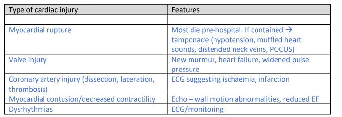
PromptRole of Troponin in blunt cardiac injury
ResponseThe use of biomarkers is unclear. Patients with haemodynamic instability and/or abnormal ECG/dysrhythmia will need echo & admission for monitoring regardless of troponin results. If there is no haemodynamic instability, no physical signs suggesting cardiac injury and no ECG abnormality an elevated troponin is non-specific & has little prognostic value. Moreover, troponin elevation occurs frequently in the setting of trauma (without cardiac injury) from catecholamine-induced stress, hypovolaemic shock with hypoperfusion/reperfusion injury, microcirculatory dysfunction etc) so and likely results in unnecessary admissions
(Nsyd 2021.1-13)
(Nsyd 2021.1-13)