CARDIOLOGY
STEMI EQUIVALENTS
PromptSgarbossa criteria - interpret ECG
ResponseSee ECG library - Be able to do this
PromptSgarbossa criteria – Describe your expected findings in the modified sgarbossa criteria for acute MI
ResponseDiagnosis of STEMI in LBBB using modified sgarbossa criteria. Any of the following is a STEMI
Any lead with >1mm concordant ST elevation (QRS and ST in same direction)
Any lead V1-V3 with >1mm concordant ST depression (QRS and ST in same direction)
Any lead with ST elevation more than 25% of a preceding S wave
New onset LBBB in a stable patient with chest pain is no longer an indication for reperfusion.
LBBB AND strong clinical suspicion is an indicator for reperfusion.
Any lead with >1mm concordant ST elevation (QRS and ST in same direction)
Any lead V1-V3 with >1mm concordant ST depression (QRS and ST in same direction)
Any lead with ST elevation more than 25% of a preceding S wave
New onset LBBB in a stable patient with chest pain is no longer an indication for reperfusion.
LBBB AND strong clinical suspicion is an indicator for reperfusion.
- PACSA
PromptDeWinter T waves
ResponseSTEMI equivalent
Upsloping ST depression V2-V5
Tall T waves in chest leads V2-V5
Slight ST elevation AVF >0.5mm
Amaal Matu – If you can fit the QRS in the T wave – always bad
Upsloping ST depression V2-V5
Tall T waves in chest leads V2-V5
Slight ST elevation AVF >0.5mm
Amaal Matu – If you can fit the QRS in the T wave – always bad
- PACSA
PromptWellens - Interpret ECG
Response Be able to do this - See ECG library
PromptWellens - Describe
ResponseECG changes suggestive of critical proximal LAD stenosis
No precordial Q waves
Normal precordial wave progression
Minimally elevated ST segments (<1mm)
Inverted or biphasic T waves V1-V6 (mainly V2-V3)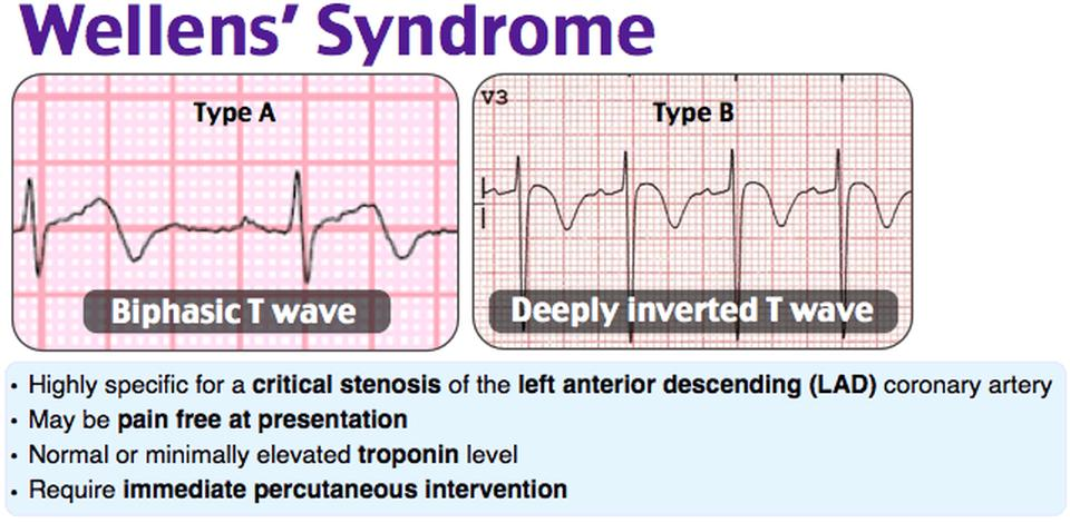
No precordial Q waves
Normal precordial wave progression
Minimally elevated ST segments (<1mm)
Inverted or biphasic T waves V1-V6 (mainly V2-V3)
- Wellens A = Biphasic
- Wellens B = Deeply inverted
PromptDifferential wellens
ResponsePE/RV strain
RBBB
HCM
Raised ICP
Normal paeds
Persistent juvenile T wave inversion
Vasospasm/cocaine
Brugada
RBBB
HCM
Raised ICP
Normal paeds
Persistent juvenile T wave inversion
Vasospasm/cocaine
Brugada
PromptMgmt Wellens
ResponseManagement as per ACS – Aspirin, Plavix/Ticag, heparin
Admit CCU monitored bed (If rural should be transferred for PCI over an admission)
Immediate PCI if redevelops symptoms
Admit CCU monitored bed (If rural should be transferred for PCI over an admission)
- Serial ECG’s to assess for development of LAD/Anterior STEMI
- GTN
- Beta blocker (metoprolol vs IV)
Immediate PCI if redevelops symptoms
PromptInferior STEMI
ResponseElevation II, III, aVF (+/- preceeded by hyperacute T waves)
Reciprocal ST depression AVL
Progressive development of Q in II, III, aVF
Concomitant RV infarction – Severe hypotension with nitrates. AVOID
RCA in 80% >> STE III>II, Reciprocal depression I, RV infarction (STE V1)
LCx 18% >> STE II=III, no reciprocal depression I, Lateral infarction – I, AVL, V5-V6 Elevation.
20% of inferior STEMI will develop 2nd or 3rd deg AV blocks. (AV node ischaemia, increased vagal tone)
- LITFL
Reciprocal ST depression AVL
Progressive development of Q in II, III, aVF
Concomitant RV infarction – Severe hypotension with nitrates. AVOID
RCA in 80% >> STE III>II, Reciprocal depression I, RV infarction (STE V1)
LCx 18% >> STE II=III, no reciprocal depression I, Lateral infarction – I, AVL, V5-V6 Elevation.
20% of inferior STEMI will develop 2nd or 3rd deg AV blocks. (AV node ischaemia, increased vagal tone)
- LITFL
PromptECG comments
ResponseSTE III>2 consistent with RV involvement – They love this. Means NO nitrates
PromptAF
Response
PromptRate vs rhythm control - Decision making
ResponseFavouring RHYTHM control
Young patient
Sevre symptoms
Short duration of symptoms
Heart failure due to AF
Mild or moderat L atrial enlargement only
Favouring RATE control
Age >65
Asymptomatic
Longstanding/persistent AF
L atrial enlargement
Young patient
Sevre symptoms
Short duration of symptoms
Heart failure due to AF
Mild or moderat L atrial enlargement only
Favouring RATE control
Age >65
Asymptomatic
Longstanding/persistent AF
L atrial enlargement
PromptList Drugs for AFib (rate/rhythm control)
ResponseBasically, pick the drug based on the side effect profile
RATE control
RATE control
- Beta blockers - Metoprolol 25 mg PO or 2.5-5mg IV – Ind: normal BP, minimal/no CCF, No Contra (eg: asthma)
- Ca blocker – Verapamil 2.5mg IV to 15 m – ind: Avoid hypotension and current CCf, avoid if on beta blocker. No beta blockade so good if Beta blocker CI (eg asthma)
- Digoxin 500 mics PO/V – ind: Low BP, Presence of CCF. Has slow onset of action
- MgSO4 – 10-20 mmol slo IV – may aide in rate control, care with hypotension
- Flecainide 2mg/kg PO/IV – Structurally normal heart if AF <48 hours
- Amiodarone 5mg/kg slow IV – If flecainide contraindicated and AF <48h. CI iodine allergy, hypotension, thyroid dysfunction, long QT
- Sotalol 40-80 mg – CI Long QT, asthma, hypotension
PromptList causes of AF
ResponseCardiac
- Htn
- Valvular disease (inc Rheumatic valvular heart disease)
- IHD/CAD
- CHF
- Cardiomyopathy
- Genetic
- Post cardiac surgery
- Sick sinus syndrome, Pre-excitation syndromes
- Pericarditis
- Hyperthyroid
- Sepsis
- EtOH – “holiday Heart”
- OSA
- COPD
- Stimulants
- PE
- Electrolyte abnormalities (Hypokalaemia, Hypomagnesaemia)
- Hypothermia = slow AF
PromptDescribe your approach to anticoagulation in AF
ResponseRisk of stroke = CHADS2VASc (Definitely Anticoagulate if M >=2, F>=3, consider if >0)
CHF
Htn
Age >65
Diabetes
Stroke (2)
Vascular disease
Age >75
Sex (female)
Risk of bleeding = HAS BLED (0-1 = anticoagulated, >=3 HIGH risk so use alternate therapy, 2 = consider. Score of 4 = 4% risk of major bleed in 100 patient years)
Htn
Abnormal liver/kidney function
Stroke
Bleeding
Labile INR
Elderly (age >65)
Drugs or alcohol
CHF
Htn
Age >65
Diabetes
Stroke (2)
Vascular disease
Age >75
Sex (female)
Risk of bleeding = HAS BLED (0-1 = anticoagulated, >=3 HIGH risk so use alternate therapy, 2 = consider. Score of 4 = 4% risk of major bleed in 100 patient years)
Htn
Abnormal liver/kidney function
Stroke
Bleeding
Labile INR
Elderly (age >65)
Drugs or alcohol
PromptIndication for Cardioveresion in AF
ResponseHaemodynamic instability
Definite onset of AF within 24-48 hours
Lack of known structural heart disease
Non chronic AF
Correctable/clear precipitant
Patient preference.
Definite onset of AF within 24-48 hours
Lack of known structural heart disease
Non chronic AF
Correctable/clear precipitant
Patient preference.
PromptSYNCOPE
ResponseLots of questions ask you to list and justify investigations for each differential. Think about that as you answer these.
PromptDefine Syncope
ResponseTransient LOC and loss of postural tone secondary to insufficient cerebral perfusion that spontaneously and completely resolves without medical intervention
PromptHistory of syncope***
ResponseRed flags:
Unheralded
Seated/supine
Exertional
FHx sudden cardiac death
Personal history/prior hx of unexplained history
Congenital cardiac disease
Unheralded
Seated/supine
Exertional
FHx sudden cardiac death
Personal history/prior hx of unexplained history
Congenital cardiac disease
PromptExamination in syncope
ResponseBP – lying and standing
Heart rate - ? dysrhythmia, bradycardia ?increased on standing
Volume status - ? dehydrated
Pallor suggesting anaemia / blood loss
PR for melaena
Presence of pacemaker
SaO2 – e.g. hypoxia in content of PE (unlikely if patient now asymptomatic)
Heart rate - ? dysrhythmia, bradycardia ?increased on standing
Volume status - ? dehydrated
Pallor suggesting anaemia / blood loss
PR for melaena
Presence of pacemaker
SaO2 – e.g. hypoxia in content of PE (unlikely if patient now asymptomatic)
PromptDDx cardiac syncope ***
ResponseComplete heart block – presence of bifascicular block on ECG in ED
VT – Hx MI, frequent non sustained VT in ED
TDP – Long QT on ECG
Severe AS – Loud harsh ejection systolic murmur with poor volume carotid pulse
HOCM – Exercise induced syncope. ECG features LVH with dagger Q waves in lateral leads
ARVC - epsilon waves on ECG, RBBB, FHx sudden cardiac death
VT – Hx MI, frequent non sustained VT in ED
TDP – Long QT on ECG
Severe AS – Loud harsh ejection systolic murmur with poor volume carotid pulse
HOCM – Exercise induced syncope. ECG features LVH with dagger Q waves in lateral leads
ARVC - epsilon waves on ECG, RBBB, FHx sudden cardiac death
PromptConcerning ECG’s in syncope (Describe what you’re looking for) ***
ResponseArrhythmia (VT, VF, rAF, etc… - ironically blatant stuff like this not commonly in answer)
Trifascicular block (3rd degree AV block) or bifascular block (risk of deterioration to 3rd degree HB)
Long QT (progression to Torsades)
Q waves – Previous MI, risk of VT
Wolf-Parkinson White - Pre-excitation/delta waves, QRS>110, PR <120. Progression to (supraventricular) reentry tachycardias
Brugada syndrome – RBBB with coved/saddleback ST elevation in V2-V3 (progression to VT/VF)
ARVD – TWI V1-V3, epsilon waves (progression to Vt/VF)
Broad QRS >120 ms - risk of VT due to structural heart disease
Deep Q/T inversion- HOCM
ECG features of severe hyper or hypokalaemia
ECG features of severe Na channel blockade
Pacemaker malfunction
Trifascicular block (3rd degree AV block) or bifascular block (risk of deterioration to 3rd degree HB)
Long QT (progression to Torsades)
Q waves – Previous MI, risk of VT
Wolf-Parkinson White - Pre-excitation/delta waves, QRS>110, PR <120. Progression to (supraventricular) reentry tachycardias
Brugada syndrome – RBBB with coved/saddleback ST elevation in V2-V3 (progression to VT/VF)
ARVD – TWI V1-V3, epsilon waves (progression to Vt/VF)
Broad QRS >120 ms - risk of VT due to structural heart disease
Deep Q/T inversion- HOCM
ECG features of severe hyper or hypokalaemia
ECG features of severe Na channel blockade
Pacemaker malfunction
PromptDDx Non cardiac syncope **
ResponseReflex (Neurally mediated) syncope
Ectopic pregnancy in young woman
- Vasovagal – sudden sensation of pain, prolonged standing. (presyncope/heralded, inciting event, post syncopal vagal sx – sweating, n+V, hx of similar)
- Situational – strain with closed glottis, micturition, defecation, swallow
- Carotid sinus – tight collar, head turning
- Hypovolaemia – blood loss/fluid loss
- Addisons
- Aortic dissection (thoracic or abdominal)
- PE
- Subclavian steal
- Sah, TIA, migraine, shy-Drager, subclavian steal
- Autonomic dysfunction
- Cardiac: Beta blocker, CCB, nitrates, ACEi, Diuretics
- Non cardiac: Antipsychotics, Antidepressants, Antiparkinsons
- Party – Cocaine, sildenafil, alcohol, poppers
- 50% in young adults
- Hypoglycaemia
- Diarrhoea/gastro
Ectopic pregnancy in young woman
PromptDDx young female with abdo pain and syncope
ResponseMUST Say ECTOPIC PREGNANCY >> positive FAST
Pericardial effusion +/- tamponade (autoimmune stuff can happen in the younger population >> bedside echo showing fluid/RA diastolic collapse
Aortic dissection (marfans in young) >> bedside echo showing intimal flap
Bedside abdo u/s showing aortic hematoma>> CT aortogram, CXR
PE >>-bedside echo, ecg, ABG, CTPA
Cardiac - MI, Dissection, Pericardial Effusion, Arrhythmia (Brady or Tachy)
Respiratory - PE
Abdominal - Ovarian Torsion, AAA (unlikely)
Neurological (CVA, SAH, Seizure)
Other - Vasovagal, Vertebral Dissection/insufficiency, carotid sinus
Pericardial effusion +/- tamponade (autoimmune stuff can happen in the younger population >> bedside echo showing fluid/RA diastolic collapse
Aortic dissection (marfans in young) >> bedside echo showing intimal flap
Bedside abdo u/s showing aortic hematoma>> CT aortogram, CXR
PE >>-bedside echo, ecg, ABG, CTPA
Cardiac - MI, Dissection, Pericardial Effusion, Arrhythmia (Brady or Tachy)
Respiratory - PE
Abdominal - Ovarian Torsion, AAA (unlikely)
Neurological (CVA, SAH, Seizure)
Other - Vasovagal, Vertebral Dissection/insufficiency, carotid sinus
PromptInvestigations for syncope (And justify)
ResponseECG – dysrhythmia, AV block, PPM dysfunction
Blood sugar - ?hypoglycaemia
Electrolytes / VBG - ?electrolyte dysfunction
Urea & creatinine - ?evidence of dehydration
Hb - ?anaemia, MCV - ?evidence of chronic blood loss (low MCV)
(TSH, BSL, BHCG) - alternative cause
Serial ECG (?dynamic changes) and continuous monitoring (monitor for arrhythmia)
Postural BP
CXR – SOB with focal examination findings eg crackles
Consider bedside Echo ?alternate cause (PE, effusion, etc although clinically unlikely)
Pacemaker check
Holter monitor – could be arranged on discharge if concerns re dysrhythmia
CT brain – if severe headache (e.g. ?SAH) or concerns that this was a seizure rather than syncope (Note CTB usually doesn’t pay in syncope questions unless trauma as result of syncope. Read Q carefully)
Blood sugar - ?hypoglycaemia
Electrolytes / VBG - ?electrolyte dysfunction
Urea & creatinine - ?evidence of dehydration
Hb - ?anaemia, MCV - ?evidence of chronic blood loss (low MCV)
(TSH, BSL, BHCG) - alternative cause
Serial ECG (?dynamic changes) and continuous monitoring (monitor for arrhythmia)
Postural BP
CXR – SOB with focal examination findings eg crackles
Consider bedside Echo ?alternate cause (PE, effusion, etc although clinically unlikely)
Pacemaker check
Holter monitor – could be arranged on discharge if concerns re dysrhythmia
CT brain – if severe headache (e.g. ?SAH) or concerns that this was a seizure rather than syncope (Note CTB usually doesn’t pay in syncope questions unless trauma as result of syncope. Read Q carefully)
PromptCTB indications in syncope
ResponseCT Head – only if signs of trauma indicating possible TBI ie signs of BOS fracture complicating fall OR lateralising neurology/signs of raised ICP
Focal neurological symptoms/signs
Persistent reduced LOC
Further unexplained seizure activity in ED
Bleeding disorder
Fever or suspected CNS infection
Past H/O intracranial pathology or malignancy
Suspected SAH
Focal neurological symptoms/signs
Persistent reduced LOC
Further unexplained seizure activity in ED
Bleeding disorder
Fever or suspected CNS infection
Past H/O intracranial pathology or malignancy
Suspected SAH
PromptScoring systems (Sanfrancisco or Chess score)***
ResponseCHESS
CHF
Haematocrit <30%
ECG abnormal
Systolic BP <90% at triage
SOB
1 q wanted 3 scoring systems listed (not described) - Canadian syncope score, San Francisco syncope rule, Sris sstratification of syncope in the ED (ROSE), Simplified Boston syncope rules, Short Term prognotis of Syncope (STePS)
CHF
Haematocrit <30%
ECG abnormal
Systolic BP <90% at triage
SOB
1 q wanted 3 scoring systems listed (not described) - Canadian syncope score, San Francisco syncope rule, Sris sstratification of syncope in the ED (ROSE), Simplified Boston syncope rules, Short Term prognotis of Syncope (STePS)
PromptClinical presentation favouring Syncope vs Seizure**
ResponseFavouring syncope
- Rapid neurologic recovery, no post ictal period
- Postural symptoms
- Absence of seizure like movements
- Pre-syncope symptoms
- No neurologic symptoms
- Hx seizures
- Persistent reduced LOC
- Further unexplained seizure activity in ED
- Bleeding disorder
- Fever or suspected CNS infection
- Past hx of intracranial pathology/malignancy
- Suspected SAH
- Time course – sudden onset/offset, brief – usually less than 2 minutes
- Description of attack – involuntary movement, incontinence, tongue biting (and location
- of tongue injury), drooling
- Amnesia
- Post ictal period – confusion, lethargy
PromptDischarge in syncope
(Q was seizure vs syncope… answer not ideal)
(Q was seizure vs syncope… answer not ideal)
ResponseNo indication for admission – ie further seizures, need for urgent treatment of any underlying cause.
Stable – no further seizures or reduced LOC
Time of day
Support at home/NOK
Follow up / further investigation as appropriate for diagnosis
Safety advice – driving, working with machinery as appropriate for diagnosis
Stable – no further seizures or reduced LOC
Time of day
Support at home/NOK
Follow up / further investigation as appropriate for diagnosis
Safety advice – driving, working with machinery as appropriate for diagnosis
PromptSafe Discharge criteria in Syncope
ResponseNo indication for admission (no treatment required for underlying cause)
Stable
Time of day
Support at home/NOK
Follow up/further investigations organised as appropriate for diagnosis
Safey advice – driving, machinery work,
Stable
Time of day
Support at home/NOK
Follow up/further investigations organised as appropriate for diagnosis
Safey advice – driving, machinery work,